The biological assays were conducted on the crude extracts to determine whether they show activity that could be used as a cosmetic ingredient in later formulations.
DPPH Antioxidant Assay
Antioxidants are compounds that help protect cells and biological materials from oxidative damage caused by free radicals and other reactive species. Free radicals can react with important molecules such as lipids, proteins and DNA, potentially contributing to cellular damage and oxidative stress. Measuring the ability of a substance to neutralise free radicals provides an indication of its potential antioxidant activity.
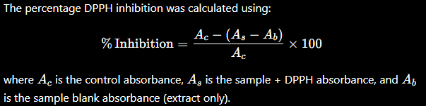
The DPPH (2,2-diphenyl-1-picrylhydrazyl) assay is a commonly used method for evaluating free-radical-scavenging activity. DPPH is a stable radical with an intense purple colour. When an antioxidant donates a hydrogen atom or electron to DPPH, the radical is reduced, resulting in a decrease in the intensity of the purple colour. This change is monitored spectrophotometrically at 517 nm, where a greater decrease in absorbance generally indicates stronger radical-scavenging activity. Ascorbic acid is commonly used as a reference antioxidant.
DPPH radical + antioxidant → reduced DPPH → decrease in absorbance
DPPH radical (violet)reduced DPPH (pale)
Experimental method
A DPPH stock solution of 1000 µM was prepared by dissolving 19.72 mg of DPPH in 50 mL, then diluted ten-fold to obtain a 100 µM working solution. The extract stock solution was prepared at 10 mg/mL by dissolving 100 mg of extract in 10 mL. Serial dilutions were prepared at 200, 100, 50, 25 and 10 µg/mL. Ascorbic acid was used as the standard antioxidant at 1 mg/mL, followed by serial dilutions of 100, 50, 25, 10 and 5 µg/mL.
For the assay, 150 µL of the 100 µM DPPH working solution was mixed with 50 µL of the respective extract dilution in the experimental wells. The control consisted of 150 µL DPPH + 50 µL ethanol, while the sample blank contained 150 µL ethanol + 50 µL extract without DPPH. The positive standard consisted of 150 µL DPPH + 50 µL ascorbic acid. Absorbance was measured at 517 nm at 0, 5, 10, 15, 20, 25, 30, 35, 40, 45, 50, 55 and 60 min, with samples shaken before each measurement.
Anti-Tyrosinase Assay
Tyrosinase is a copper-containing enzyme that plays an important role in melanin production, particularly by catalysing the oxidation of L-tyrosine and L-DOPA during melanogenesis. Excessive tyrosinase activity can contribute to increased melanin formation and skin hyperpigmentation. Inhibiting tyrosinase is therefore of interest in cosmetic and pharmaceutical products aimed at controlling melanin production.
The L-DOPA-based tyrosinase inhibition assay determines whether an extract can reduce tyrosinase activity. In the assay, tyrosinase converts L-DOPA into coloured oxidation products, with the resulting change in absorbance monitored spectrophotometrically. If an extract contains compounds capable of inhibiting tyrosinase, less coloured product is formed. Kojic acid is commonly used as a positive control.
tyrosinase + L-DOPA → coloured product
Experimental method
Anti-tyrosinase activity was evaluated in 96-well microplates using mushroom tyrosinase as the enzyme and L-DOPA as the substrate, with kojic acid used as the positive control. Mushroom tyrosinase (100 U/mL) was prepared in PBS (pH 6.8) by dissolving 1 mg of enzyme (1000 U) in 10 mL PBS. L-DOPA (10 mM) was prepared by dissolving 19.72 mg in 10 mL PBS. A 2 mM L-tyrosine solution was prepared by dissolving 3.62 mg in 10 mL PBS.
For the assay, 120 µL PBS, 20 µL tyrosinase and 20 µL of the respective extract were added to each test well and allowed to pre-incubate for 10 min. The enzymatic reaction was initiated by adding 40 µL of 10 mM L-DOPA, giving a final reaction volume of 200 µL. Kojic acid was tested under the same conditions. The plate was incubated at 35 °C, and absorbance was measured at 475 nm at 5-min intervals for 30 min.
The negative control contained 120 µL PBS, 20 µL tyrosinase, 20 µL solvent (ethanol) and 40 µL L-DOPA, while the solvent-free control used water instead of ethanol. A reagent blank containing 160 µL PBS and 40 µL L-DOPA, without enzyme or extract, accounted for the absorbance of the substrate. Sample blanks were prepared by combining 160 µL PBS, 20 µL extract and 20 µL L-DOPA, without tyrosinase, to correct for the intrinsic absorbance of the extracts.
Anti-Elastase Assay
Elastase is a proteolytic enzyme that hydrolyses elastin and other elastin-related proteins, contributing to the breakdown of structural components that help maintain skin firmness and elasticity. Excessive elastase activity has been associated with degradation of the extracellular matrix and processes related to skin ageing. In cosmetic and skincare research, inhibition of elastase is investigated as a potential approach for preserving skin structure and reducing the effects associated with loss of elasticity. In this assay, elastase activity was evaluated using a synthetic substrate that releases a coloured product upon enzymatic hydrolysis.
enzyme + substrate + inhibitor → reduced reaction
Experimental method
Anti-elastase activity was determined using a colorimetric assay based on the hydrolysis of N-succinyl-Ala-Ala-Ala-p-nitroanilide (SANA) by porcine pancreatic elastase (PPE). The release of p-nitroaniline was monitored spectrophotometrically at 410 nm, where a reduction in absorbance indicated inhibition of elastase activity.
A 0.1 M Tris-HCl buffer (pH 8.0) was prepared by dissolving 1.2114 g Tris in approximately 80 mL distilled water, adjusting pH to 8.0 with HCl and making the volume up to 100 mL. PPE was prepared as a 10 U/mL working solution by dissolving 6.25 mg enzyme in 2.5 mL Tris-HCl buffer. The SANA substrate was prepared at 10 mM by dissolving 9.81 mg SANA in 2.5 mL Tris-HCl buffer, with a small volume of DMSO used to facilitate dissolution.
The test extracts were prepared at 20,000 µg/mL by dissolving 200 mg of extract in 10 mL ethanol. Serial two-fold dilutions were prepared to obtain concentrations of 10,000, 5,000, 2,500, 1,250, 625, 312.5, 156.25 and 78.125 µg/mL. Ursolic acid was used as the positive control at an initial 10,000 µg/mL by dissolving 100 mg in 10 mL DMSO, followed by dilution to the required concentrations.
The assay was performed in a 96-well microplate using a final reaction volume of 200 µL per well. Each reaction well contained 140 µL Tris-HCl buffer, 20 µL of the respective extract or ursolic acid solution, and 20 µL of PPE. The plate was incubated at room temperature for 15 min to allow interaction between enzyme and test samples. The reaction was initiated by addition of 20 µL of 10 mM SANA substrate, after which the plate was incubated in the dark for 30 min. Absorbance was measured at 410 nm at 5-min intervals.
Solvent controls were included to account for any effects of the extraction solvents on enzyme activity. The ethanol control contained 160 µL buffer, 20 µL PPE, 20 µL ethanol and 20 µL SANA, while the DMSO control contained 160 µL buffer, 20 µL PPE, 20 µL DMSO and 20 µL SANA. A reagent blank containing 180 µL buffer and 20 µL SANA was used to account for background absorbance. Sample blanks containing 180 µL buffer and 20 µL test sample, without enzyme, were included to correct for the intrinsic absorbance of the extracts.
An instrument reading is never automatically the final result. Sample, solvent, reagent and plate background all contribute to the signal — so every value moved through correction and calculation before it became a graph, and only then an interpretation. Some biological work images were not taken due to the hygiene requirements of these sensitive experiments.